How to help surgeons perform breast cancer surgery more successfully
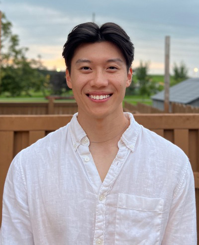
 Lawrence Yip, a PhD candidate in the Department of Medical Biophysics at Western University, works at St. Joseph’s Hospital in Dr. Jeffrey Carson’s lab developing a new imaging technology to help surgeons treat breast cancer. In collaboration with his lab colleagues, Lawrence seeks to build a portable system that will be reliable and inexpensive.
Medical Biophysics is an interdisciplinary field where the principles of the physical sciences are employed to define and explore living things, often for the purposes of medical application. “As an interdisciplinary department, Medical Biophysics is a ‘big mix’”, says Lawrence.“ Collaboration is the key factor here because we involve a lot of transdisciplinary research from medical imaging, cell biology, physiology, kinesiology and many other areas to apply them directly to patient care.”
What Lawrence loves most in his academic life now is the focus on translational research and the chance provided by his lab to “build something from scratch”. “Even as a kid I enjoyed designing little things. My dad, an electrical engineer, inspired me to build something from common materials and taught me how to create things from the ground up. I find all those skills extremely helpful for what I am focusing on in my lab.”
The portable device that Lawrence currently is building will help surgeons perform cancer surgery more successfully. The technology, which is called 3D photoacoustic tomography (PAT), will be able to improve outcomes for cancer patients who need surgery. Especially promising is the use of this device in treating breast cancer.
At present, there is an option for some breast cancer patients to undergo a lumpectomy, which is a breast-conserving surgical technique where only tumor(s) are removed (unlike a mastectomy, in which a surgeon removes the entire breast). In a lumpectomy, the tumour is excised with a margin of healthy tissue ranging from a 2-mm to a 10-mm width.
“The key thing for the surgeon is to remove as much tumor tissue as is possible. If a substantial mass of tumor is left in the breast, it will grow back in to another tumor. To ensure complete tumour removal, the surgeons take a layer of healthy tissue right around of the whole tumor. A margin of tissue should be clean, that is, not have any cancer cells. Clear margins are the standard for lumpectomy surgery.”
 However, sometimes additional surgery may be required after a lumpectomy. As Lawrence explains, detection of the margin can be difficult because current techniques used to guide excision are often insufficient. “We need to wait for the pathology report. After the surgery is done, the pathologist examines the excised tumor to make sure that its margins have no cancer cells. It takes time. Unfortunately, current lumpectomy re-excision rates average 25% of the time when pathology finds a tumour in the margin after surgery.”
The technique Lawrence is developing will provide a way to see the margin in the operating room. “PAT is a hybrid imaging technique that combines the strengths of ultrasound and optical imaging while using non-invasive laser light. This technique is quite novel. It is around 20 years now in active research and is not yet a clinical standard. Ultrasound looks at the differences in the density of materials by using sound, while light imaging can look at very specific substances. Using light, you can target various things just by changing what color light you use. With this method, 3D images with high contrast, high resolution and excellent penetration depth can be achieved while maintaining a low cost.”
What does this system look like and how does it work? “Since PAT is a portable system, it is not as big as, say, an MRI machine. PAT can be wheeled down to any operating room or wherever we need to scan what we need. The workflow with PAT will be as follows: the surgeon takes out the tumor, brings it to the PAT system, scans the material and gets the image right away, which shows if the margins are cancer-free or if the tissue must be excised more. The entire process will take 5 -10 min while the patient is in the operating room. So, this system will help surgeons ensure that complete tumour removal is achieved on the first try. No re-excision will be necessary.”
Lawrence hopes to use the system in the operating room shortly to define its applicability with real breast tumours and to compare its performance with previous generation PAT systems. “The hardest thing in my field is when you finish something, and you think it should work, but it doesn’t. You should step back and try something different. Persistence is highly important in research. All of us in the lab work hard and hope that our systems will work effectively from the first try and will be used in clinical practice soon.”
Support researchers like Lawrence Yip and others by considering a donation to the Breast Cancer Society of Canada. Find out how you can help fund life-saving research, visit bcsc.ca/donate
Lawrence Yip’s story was transcribed from interviews conducted by BCSC volunteer Natalia Mukhina – Health journalist, reporter and cancer research advocate
 Natalia Mukhina, MA in Health Studies, is a health journalist, reporter and cancer research advocate with a special focus on breast cancer. She is blogging on the up-to-date diagnostic and treatment opportunities, pharmaceutical developments, clinical trials, research methods, and medical advancements in breast cancer. Natalia participated in numerous breast cancer conferences including 18th Patient Advocate Program at 38th San Antonio Breast Cancer Symposium. She is a member of The Association of Health Care Journalists.

